
Insane in the membrane....
If you're at all squeemish, you may care to look away now.
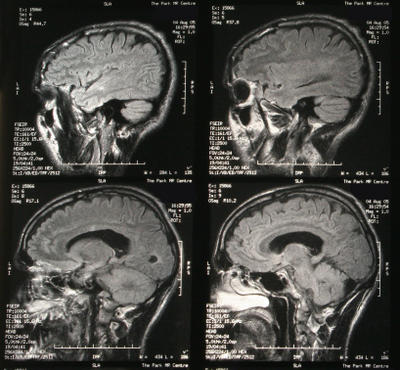
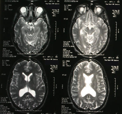
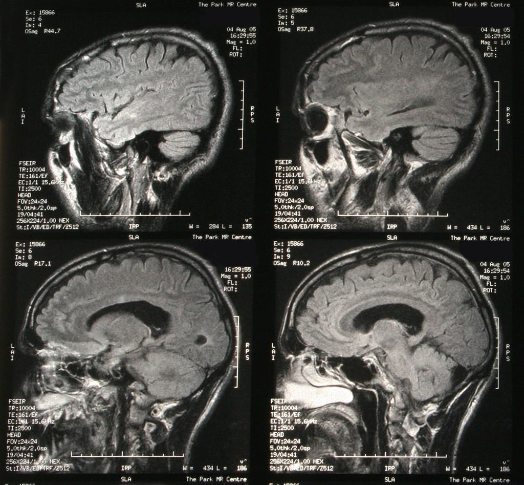
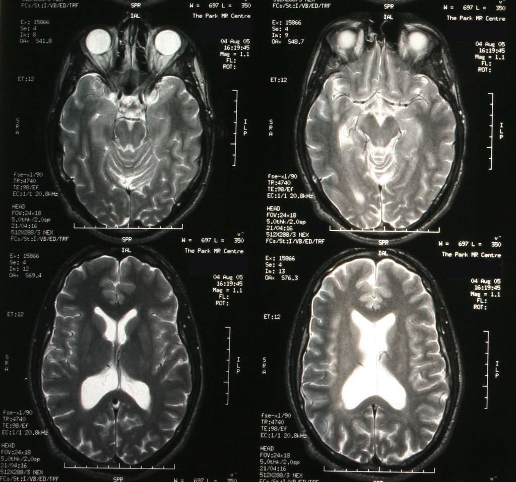
Allow me to present for your viewing pleasure...... my brain.
(Feel free to click on an image if you want a close-up....)
I ask you - what other blogger gives you more? Could you ask for a greater insight into the innermost workings of my mind?
Samuel Pepys eat your heart out.
And yes, now you mention it, I do look a little bit like a fish in some of these shots....
(**respect due to the Eye In The Sky for these quality photos of the original scans....**)
posted by swisslet @ 00:29
![]()
![]()


14 Comments:
At 4:24 am, HistoryGeek said…
I particular like the top left of the bottom 4...it reminds me of a trilobite.
So do we know what it all means? Or do you have to wait to talk to another MD?
At 6:29 am, Aravis said…
The bottom set reminds me of rorschack (sp?) tests. The top two of this set reminds me of screaming aliens. Is that by any chance what's causing your problem? I wouldn't have thought so, but then I'm not a doctor. *G*
Seriously though, hope that these revealed that you are well.
At 8:42 am, Ali said…
>screaming aliens
I concur with your diagnosis aravis, although the bottom two are definitely pancakes. That might be causing headaches.
Also, is that an earworm I see in one of the shots?
Bearing in mind I haven't a clue what I'm on about, other than that it all looks nice a healthy to me. No shrinkage or atrophy, and no evidence of plaques or lesions. (I almost sounded convincing then, didn't I?)
When do you hear back?
At 9:47 am, swisslet said…
well, here's what the neurologist said in full:
Brain: Coronal T1 and T2 sagittal FLAIR
There is no evidence of a space occupying lesion. There is a mild enlargement of the posterior bodies and occipital horns of the lateral ventricles with a slightly colpocephalic appearance. Areas of ill defined high signal change on FLAIR and T2 imaging are noted adjacent to the bodies of th elateral ventricles bilaterally and a small subcortical high signal lesion is noted involving the right middle temporal gyrus. There is no definite involvement of the corpus callosum or posterior fossa structures.
Cervical Spine: Sagital T1 T2 axial T2* sagital and axial T1 plus Gadolinium.
vertebral height and alignment are maintained. Canal dimensions are generous. There is a discreet oval high signal lesion located ventrally in the cord at C3 level causing mild cord expansion. This is poorly visualised on T1 imaging but shows evidence of marginal enhancement on the post contrast images. Axial images show the lesion lies on the left side of the cord at the C3 level. No other cervical cord lesions are identified.
Thoracic cord: Sagital T1, T2 and T1 plus Gadolinium.
Vertebral height and alignment are maintained. Canal dimensions are generous. There is no evidence of extrinsic compression of the thoracic cord, or of an intermedullary lesion.
Conclusion: The combination of the definite left ventral cord lesion at C3 and the subappenydymal lesions on the brain imaging is rather suggestive od demyleination.
----
And what does that mean? Well, my neurologist actually disagreed with the hospital neurologist that wrote the report, and said he couldn't see any lesions in my brain, but only on my cervical spinal cord. This is good - I don't want lots of lesions, as that way MS lies. I have a single lesion that seems to be on the way down. It's probably caused by a virus and may take about 3 months to go away and they may never know what caused it. It may mark my spinal cord permanently and leave a physical limit that wasn't there before, albeit one that won't affect me in my every day life, but might come into play when I really push myself physically and my nervous system will just say "NO". Ho hum. Back in 3 months time for a follow up appointment, and in the meantime just wait.
It's if it comes back that I really have to start to worry.
The colleague of mine who had similar symptoms to me at the same time has just been diagnosed with MS, so I'm currently feeling that there but for the grace of God go I....
So in short: I'm okay, I think.
ST
At 10:14 am, Damo said…
Ooooh! Shiny.
At 10:14 am, Damo said…
And... glad it all looks OK.
At 12:25 pm, John McClure said…
Check out the big brain on Brett!
I can't believe no one has mentioned what looks like a gaping hole in the middle of your brain. You'd think they'd train these doctors to spot stuff like that.
At 2:37 pm, Ali said…
>So in short: I'm okay, I think.
Glad to hear that. You do realise your brain slice pictures gave me bad dreams last night. No, really, they did. See LJ for details.
Glad you're okay. My old friend. LOL!
At 5:00 pm, HistoryGeek said…
I'm glad things look okay.
At 5:53 pm, red one said…
The parts you have provided seem fine, but you've missed out the instructions on how to join the bits together - attach tab A to tab C etc - so we can make our own Swiss Toni brains.
Seriously though, glad it seems to be OK. And you are a Proper Blogger posting full details of your brain like that. Not to mention admirably matter of fact about it though it must all have been rather scary.
red
At 8:46 pm, Mark said…
it looks like some weird deep sea angelfish to me. that could explain plenty...
At 3:58 pm, swisslet said…
I've actually got a whole folder full of these things now...100s of them (they really give you your money's worth, apparently). Seemingly endless cross-sections of my head, neck and spinal cord.
I took them home this weekend to show my dad (who is a doctor) and he had a look at them, and after remarking that "we didn't have these in my day", his single useful observation was that you could see that my nose wasn't straight because the sinus channels run off wonkily.... not that you need an MRI scan to tell that, just a pair of eyes.
He couldn't see the lesion, which just goes to show how good the people who do spot them are and also why the two neurologists disagree over how many I've got.
I'd post more, but I've kind of got MRI fatigue, and I reckon these are the best ones anyway. It makes me feel a bit weird looking at them, actually....
ST
At 5:29 pm, adem said…
I just popped by to say Hi. Cool scans by the way....I want some! (I reckon you could make a mint selling them on ebay)
At 6:57 am, Me said…
Glad you're ok, ST! It must be very strange looking at your own brain. It's strange enough looking at someone else's.
Mike
Post a Comment
<< Home